TGCT Support
  • Home
  • About TGCT
    • Causes of TGCT
    • Types of TGCT
    • Tumor Location
    • TGCT in Pediatrics
    • Diagnosis >
      • Biopsy
      • Imaging Features
  • Treatment
    • Surgery >
      • Surgery Guide
      • Recovery
    • Medications >
      • Turalio >
        • Recommended Dosing
      • Romvimza
      • Imatinib
      • Common Side Effect of TGCT Drugs
    • Clinical Trials >
      • AMB-05X
      • Pimicotinib
      • Emactuzumab
      • Cabiralizumab
      • Lacnotuzumab
    • Radiation
  • Get Support
    • Join the Community >
      • Volunteer
      • Support Groups
    • TGCT Specialists
    • Consensus Guideline
    • Our Research
    • Nutrition >
      • TGCT Cookbook
    • Pain Management
    • Mental Health >
      • Find Mental Health Help
    • Glossary of Terms
    • Educational Materials & Videos >
      • Physical Therapy
    • Webinars
    • FAQs
  • About Us
    • Meet the Team
    • Medical Advisory Board
    • Partners >
      • TGCT Support France
    • Contact Us
    • Our Store
  • News
    • Events
    • Patient Stories
  • DONATE
    • GEM Program

imaging features

Imaging Techniques

TGCT can be diagnosed through MRI and/or biopsy. Often, TGCT presents with some unique imaging features that can distinguish it from other diseases. However, commonly, ruling out other more common diseases (differential diagnosis) is still required. Under a microscope, localized TGCT and diffuse TGCT look identical. However, these subtypes appear different on MRI images. 

It is estimated that in 33% of diffuse TGCT patients, bone abnormalities are observed(1). Other studies suggest that one in five patients (20%) have secondary joint degeneration (osteoarthritis) from TGCT (2). Additionally, cysts in non-weight bearing regions, such as Baker cysts, commonly accompany TGCT of the knee due to pooling of fluid in the back of the knee. 

Consult with your radiologist and multi-disciplinary healthcare team to interpret your results.

MRI features

Magnetic resonance imaging (MRI) allows for soft tissue to be visualized in the most clear and optimal way. A MRI is an imaging technique that utilizes a strong magnetic field and radio waves to generate an image that cannot be seen well on x-rays. MRI does not use radiation, instead, it relies on atoms in your body to align with the moving magnets in the scanner. MRI is an important tool to determine TGCT subtype, as described in Diagnosis. MRI images are broken down into components: T1, T2, and GRE. Each component tells the radiologist something about anatomical structures in that area.

Our body is made up of protons, neutrons, and electrons. The MRI creates an image by aligning protons with the magnetic field created by magnets moving in the scanner. T1 and T2 describe the relaxation time of the patients' protons under the MRI after different radiographic frequencies are pulsed (different ways the magnet can move in the machine). Different structures in our body create different relaxation times. For example, fat and bone have different relaxation times and show up differently on the MRI. These sequences are used together to understand the health of the joint.

For instance, T2 weighted imaging is typically used to image chronic and progressive diseases compared to T1 weighted (3). T2 weighted imaging is often compared to T1 to identify tumors or masses. Gradient recalled echo (GRE) is a type of spinning proton, leading to specific signals. For example, metal artifacts create low GREs. Because TGCT has hemosiderin, an iron-storing compound, radiologists can sometimes see a low GRE signal, low T1, and low T2 interval signal. 

MRI results can be complex, consult with your radiologist and healthcare provider to go over MRI reports.

Localized

  • Localized TGCT often appears well-defined and discrete with clear boundaries 
  • Often stays within the joint space (e.g., Hoffa pad of knee)


Diffuse

  • ​Diffuse often appears wide spread and may be in several areas of a joint and may encompass swelling (effusions)​
  • ​Often extends outside the joint space and into neighboring structures
  • Hemosiderin deposits are seen as enhancements in the MRI, possibly giving a low signal on GRE sequences
  • "Blooming artifacts" are observed in 86% of diffuse patients and 27% of localized (4), this means the tumor may look bigger than it actually is.
  • Intermediate to weak signal of T2 distinguishes TGCT from ganglion cysts, nerve-sheath tumors, and synovial sarcoma(5).
  • However, other diseases do cause weak to intermediate signal on MRI, meaning the doctor should assess your symptoms along with the imaging
Picture
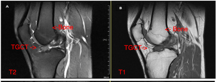
In A, T2 was used. The TGCT appears lighter than the knee bone. In B, T1 was used where the TGCT is darker than the neighboring bone. Loiy Alkhatibm, Ralf Kraus, Scott, Sigman Ulrich Stahl, Loiy Alkhatibm. 2020. Tenosynovial Giant Cell Tumor in the Tibial Attachment of the Anterior Cruciate Ligament: A Case Report

References

  1. Gouin F, Noailles T. Localized and diffuse forms of tenosynovial giant cell tumor (formerly giant cell tumor of the tendon sheath and pigmented villonodular synovitis). Orthop Traumatol Surg Res. 2017;103(1S):S91-S97. doi:10.1016/j.otsr.2016.11.002
  2. Spierenburg G, Staals E, et al. Active surveillance of diffuse-type tenosynovial giant cell tumors: A retrospective, multicenter cohort study. European Journal of Surgical Oncology. 2024
  3. Myers BW, Masi AT. Pigmented villonodular synovitis and tenosynovitis: a clinical epidemiologic study of 166 cases and literature review. Medicine (Baltimore). 1980;59(3):223-238.
  4. Julia Crim, Samantha Dryoff, James Stensby, Andrea Evenski, & Lester Layfield. Limited usefulness of classic MR findings in the diagnosis of tenosynovial giant cell tumor. Skeletal Radiology. 2021.10.1007/s00256-020-03694-4
  5. Ding Y, Griffin JE, Raghavan M, Xu H, Henderson-Jackson E, Bui MM. Tenosynovial giant cell tumors lacking giant cells: report of diagnostic pitfalls. Ann Clin Lab Sci. 2014;44(2):222-227.
Ⓒ TGCT Support
Updated 2025

TGCT SUPPORT IS A PROGRAM OF THE LIFE RAFT GROUP

Picture
155 US Highway 46, Suite 202                      Wayne, NJ 07470                      [email protected]