TGCT Support
  • Home
  • About TGCT
    • Causes of TGCT
    • Types of TGCT
    • Tumor Location
    • TGCT in Pediatrics
    • Diagnosis >
      • Biopsy
      • Imaging Features
  • Treatment
    • Surgery >
      • Recovery >
        • Surgery Guide
    • Medications >
      • Turalio >
        • Recommended Dosing
      • Romvimza
      • Imatinib
      • Common Side Effect of TGCT Drugs
    • Clinical Trials >
      • AMB-05X
      • Pimicotinib
      • Emactuzumab
      • Cabiralizumab
      • Lacnotuzumab
    • Radiation
  • Get Support
    • Join the Community >
      • Volunteer
      • Support Groups
    • TGCT Specialists
    • Consensus Guideline
    • Our Research
    • Nutrition
    • Pain Management
    • Mental Health >
      • Find Mental Health Help
    • Glossary of Terms
    • Educational Materials & Videos
    • Webinars
    • FAQs
  • News
    • Events
    • Patient Stories
  • About Us
    • Meet the Team
    • Medical Advisory Board
    • Partners >
      • TGCT Support France
    • Contact Us
    • Our Store
  • DONATE
    • GEM Program

types of TGCT

Disease characteristics are based on disease classification

The patient experience can vary drastically depending on the type of TGCT and other disease characteristics, like tumor location and prior recurrences. MRI is the primary diagnostic technique used to confirm the diagnosis of TGCT, pre-operatively (1). Additionally, it is the primary tool used to confirm recurrence following TGCT treatment. Pre-operative biopsy can be used to verify diagnosis. 

​There are two subtypes, localized/nodular and diffuse. The subtypes of TGCT can influence treatment options and disease recurrence risk (2). Under a microscope, these two subtypes look the same. However, upon MRI, differences can be determined based on tumor behavior (3). Special considerations should be taken with both forms, however, a multi-disciplinary approach may be required for those with diffuse, recurrent, or inoperable disease (4,5). ​

​Localized/nodular

Diffuse 

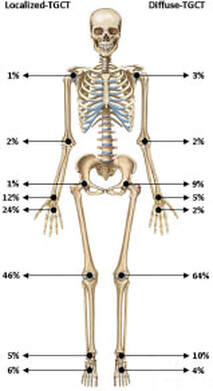
The localized form of TGCT is the most prevalent, with 85-90% of the TGCT diagnoses (6)
​
  • Tends to impact smaller joints, with the knee being most common (~46%) (7)
 
  • ​High predominance in the hands and fingers, as well as other locations, like the wrist, elbow and hip (8)
 
  • Single, well-defined tumor with a distinct border or generally encapsulated (8)

  • May stay in the joint (intra-articular) or extend out of the joint (extra-articular) (9)
 
  • Tumors typically are small, ± 2cm (8)
 
  • Often does not lead to surrounding joint damage and bone erosion (8)
​
  • Low recurrence rate (~9-15%) (6)
Picture
Mastboom MJL, Verspoor FGM, Verschoor AJ, et al. Higher incidence rates than previously known in tenosynovial giant cell tumors. Acta Orthop. 2017.

Diffuse

The diffuse form of TGCT is less common, and 2.6 times more likely to recur (7)

  • Tends to impact larger joints, with the knee being the most common (~64-75%) (10 ,11)
 
  • Can occur in other locations, like the hip (~9%) and ankle (~10%) (10)
 
  • Multiple tumors with no distinct boundary (10)
 
  • Tumors may stay in the joint (intra-articular) or extend out of the joint (extra-articular), higher likelihood of extra-articular tumors (10)
 
  • Tumors typically are large, > 5cm (10)
​
  • Can lead to local destruction of tendons, joints, surrounding bone, and infiltrate the joint capsule and surrounding muscle (12)
 
  • High recurrence rate (50-70%) (13)

Prevalence

Due to the rarity of TGCT, getting concise epidemiological information can be challenging. Prevalence among sexes and age groups can differ based on study demographics. TGCT commonly occurs between the 3rd and 5th decade of life, in both forms (10). Some studies have found that the diffuse form has a higher prevalence among younger people, 25-40 years of age (14). TGCT, in both forms, can occur in any age group.

Localized/nodular

Diffuse

  • Some studies have shown a female:male ratio of 1.5:1 (7)

  • Often seen in individuals 30-50 years old, but can vary widely (7)
 
  • Globally, it is estimated that there are 39 cases per million of localized TGCT (7)
​
  • In the United States, 9.2 cases per million are estimated to be localized TGCT (15)

Diffuse

  • No sex predilection shown (15)

  • Often seen in younger adults, < 40 years old (14)

  • Globally, it is estimated that there are 4 cases per million of diffuse TGCT (7)
​
  • In the United States, 1.8 cases per million are estimated to be diffuse TGCT (15)

Symptoms

Symptoms can vary based on many individual patient characteristics and disease features. Many of the symptoms experienced are felt by both localized and diffuse TGCT patients.

Localized/Nodular

Diffuse

  • Symptoms commonly include swelling, locking, popping, and pain (16)​
​
  • May be painless, but pain may develop with tumor growth (8)
​
  • Range of motion may be limited (14)
Picture

Diffuse

  • Symptoms commonly include swelling, stiffness, instability, pain, locking, popping, and limited range of motion (15)
​
  • Pain may develop with tumor growth. Tumor growth can lead to osteoarthritis, cartilage damage, and bone erosion (18)​
Picture

Tumor Progression

Tumor progression depends on the individual. Some tumors progress over time, some remain stable, and some grow rapidly. For some asymptomatic patients, monitoring growth is preferred over immediate medical intervention. For others, immediate surgery is done. Often, patients take 3-4 years to be diagnosed accurately due to high misdiagnosis rate and delay in seeking medical consultation.

Major Differential Diagnoses

Synovial Chondromatosis
Sport injuries such as labrum and meniscus tears
Osteosarcoma and synovial sarcoma
Fibromas
Ganglion Cysts
Xanthomas

References

 1.        Wang C, Song RR, Kuang PD, Wang LH, Zhang MM. Giant cell tumor of the tendon sheath: Magnetic resonance imaging findings
​in 38 patients. Oncol Lett. 2017. doi:10.3892/ol.2017.6011
2.      Van Der Heijden L, Gibbons CLMH, Dijkstra PDS, et al. The management of diffuse-type giant cell tumour (pigmented villonodular synovitis) and giant cell tumour of tendon sheath (nodular tenosynovitis). J Bone Jt Surg - Ser B. 2012. doi:10.1302/0301-620X.94B7.28927
3.       Murphey MD, Rhee JH, Lewis RB, Fanburg-Smith JC, Flemming DJ, Walker EA. Pigmented Villonodular Synovitis: Radiologic-Pathologic Correlation. RadioGraphics. 2008. doi:10.1148/rg.285085134
4.      Bernthal NM, Ishmael CR, Burke ZDC. Management of Pigmented Villonodular Synovitis (PVNS): an Orthopedic Surgeon’s Perspective. Curr Oncol Rep. 2020. doi:10.1007/s11912-020-00926-7
5.     Van Der Heijden L, Gibbons CLMH, Hassan AB, et al. A multidisciplinary approach to giant cell tumors of tendon sheath and synovium - A critical appraisal of literature and treatment proposal. J Surg Oncol. 2013. doi:10.1002/jso.23220
6.     
Anderson WJ, Doyle LA. Updates from the 2020 World Health Organization Classification of Soft Tissue and Bone Tumours. Histopathology. 2021. doi:10.1111/his.14265
​7.     Mastboom MJL, Verspoor FGM, Verschoor AJ, et al. Higher incidence rates than previously known in tenosynovial giant cell tumors. Acta Orthop. 2017.
​8.       
Nagase M, Araki A, Ishikawa N, et al. Tenosynovial Giant Cell Tumor, Localized Type With Extensive Chondroid Metaplasia: A Case Report With Immunohistochemical and Molecular Genetic Analysis. Int J Surg Pathol. 2020. doi:10.1177/1066896919889672
9.        Ma X, Shi G, Xia C, Liu H, He J, Jin W. Pigmented villonodular synovitis: A retrospective study of seventy five cases (eighty one joints). Int Orthop. 2013. doi:10.1007/s00264-013-1858-9
10.     
Righi A, Gambarotti M, Sbaraglia M, et al. Metastasizing tenosynovial giant cell tumour, diffuse type/pigmented villonodular synovitis. Clin Sarcoma Res. 2015. doi:10.1186/s13569-015-0030-2
11.      
Ottaviani S, Ayral X, Dougados M, Gossec L. Pigmented Villonodular Synovitis: A Retrospective Single-Center Study of 122 Cases and Review of the Literature. Semin Arthritis Rheum. 2011. doi:10.1016/j.semarthrit.2010.07.005
12.      
Giustini N, Bernthal NM, Bukata S V., Singh AS. Tenosynovial giant cell tumor: case report of a patient effectively treated with pexidartinib (PLX3397) and review of the literature. Clin Sarcoma Res. 2018. doi:10.1186/s13569-018-0101-2
13.       
Ehrenstein V, Andersen SL, Qazi I, et al. Tenosynovial giant cell tumor: Incidence, prevalence, patient characteristics, and recurrence. A registry-based cohort study in Denmark. J Rheumatol. 2017. doi:10.3899/jrheum.160816
​14.       National Organization for Rare Disorders. Tenosynovial Giant Cell Tumor. NORD. 2017. https://rarediseases.org/rare-diseases/tenosynovial-giant-cell-tumor/.
15.      Giustini N, Bernthal NM, Bukata S V., Singh AS. Tenosynovial giant cell tumor: case report of a patient effectively treated with pexidartinib (PLX3397) and review of the literature. Clin Sarcoma Res. 2018. doi:10.1186/s13569-018-0101-2
16.       
Rao AS, Vigorita VJ. Pigmented villonodular synovitis (giant-cell tumor of the tendon sheath and synovial membrane). A review of eighty-one cases. J Bone Jt Surg - Ser A. 1984. doi:10.2106/00004623-198466010-00012
17.       
Myers BW, Masi AT, Feigenbaum SL. Pigmented villonodular synovitis and tenosynovitis: A clinical epidemiologic study of 166 cases and literature review. Med (United States). 1980. doi:10.1097/00005792-198005000-00004
18.       
Gelhorn HL, Tong S, McQuarrie K, et al. Patient-reported Symptoms of Tenosynovial Giant Cell Tumors. Clin Ther. 2016. doi:10.1016/j.clinthera.2016.03.008
Ⓒ TGCT Support
Updated 2022

TGCT SUPPORT IS A PROGRAM OF THE LIFE RAFT GROUP

Picture
155 US Highway 46, Suite 202                      Wayne, NJ 07470                      [email protected]