A biopsy is a procedure where a tissue sample is removed from the patient. Upon removal of the tissue sample, the specimen can be studied under a microscope to determine the biology of the cells. Pathologists use biopsies to determine the behavior of the cell, the biology of the cell, and what type of disease the patient has. In the case of TGCT, biopsy is often necessary to confirm diagnosis when the diagnosis is unclear.
There are two main ways biopsies are done, Fine-Needle Aspiration or during a surgical procedure.
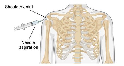
FINE-NEEDLE ASPIRATION
During fine-needle aspiration, the region the tumor is located and the surrounding region is numbed using a local analgesic. A thin needle is inserted into the tissue of interest allowing a small portion to be removed. This type of biopsy can be done in a hospital or at a doctor's office. This is considered a minimally invasive biopsy, however, there is a small risk of infection and bleeding at the needle insertion site.
Surgical biopsy
During surgery, a tissue sample can be removed allowing a pathologist to evaluate the tumor present. This is considered more invasive and generally is done in a hospital or outpatient facility. This may require the patient be fully sedated under general anesthetics. With the high accuracy of the fine-needle aspiration, surgical biopsies are less common.